
The virtual microscope
Synchrotron-based computed tomography
Synchrotron radiation is the best and brightest X-ray radiation. It has undergone considerable development in recent decades. The first description of synchrotron radiation dates back to 1947 and since then interest in it has grown, especially in physics and research with solids. The last few decades have seen new possibilities in research and diagnostics. The principle of synchrotron radiation is based on the movement of electrons. If the electrons move fast enough, they emit energy that is greater than that of X-ray waves. The electrons are introduced into the booster synchrotron and accelerated to a high speed. In the process, they are brought up to 6 giga-electron volts (GeV).
When the beam changes, it loses energy. This energy is used as synchrotron radiation to take pictures. Special magnets ensure that the radiation is coherent and bright. It is therefore as good as modern laser radiation. Synchrotron radiation is hundreds of billions of times brighter than an ordinary X-ray source, as used in medical imaging. This means that you can see better than with a normal computer tomography. Most conventional, clinically used imaging methods use the effect that X-rays are attenuated when they penetrate the tissue. In the hierarchical phase contrast tomography procedure, attenuation effects are also used. The phase shifts of electromagnetic radiation are converted into intensity fluctuations. These are recorded by the detector and reconstructed in three dimensions with high edge definition. In 2020, the ESRF was upgraded to a “fourth generation” X-ray source. This made “hierarchical” phase-contrast CT (HiP-CT) possible.

HiP-CT
The Hierarchical Phase Contrast Tomography (HiP-CT) method also makes use of attenuation effects. The phase shifts of electromagnetic radiation are converted into intensity fluctuations. These are recorded by the detector and reconstructed in three dimensions with high edge sharpness. In 2020, the ESRF was upgraded to a “fourth generation” X-ray source. This made “hierarchical” phase contrast CT (HiP-CT) possible.
All data are generated from ESRF beamtimes 1290 and 1389 and are visualized by using Siemens Healthineers Cinematic Rendering software

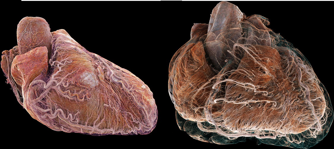
3D cinematic rendering of Hierarchicalphase-contrast tomography (HiP-CT) scans of heart tissue from autopsies.
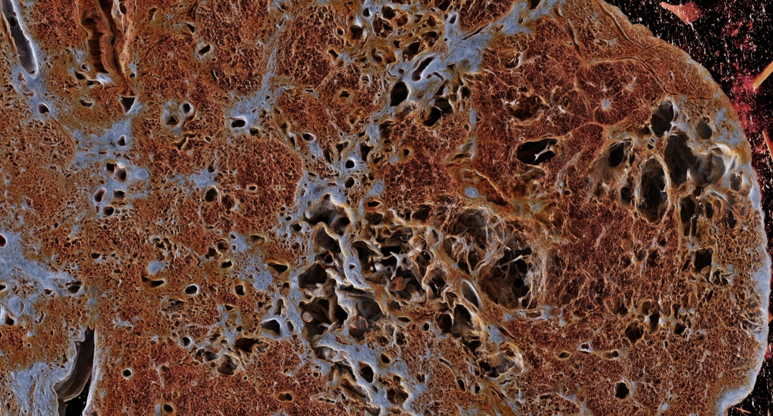
3D cinematic rendering of Hierarchical phase-contrast tomography (HiP-CT) depicting lung tissue with fibrotic remodeling and ectatic airways.
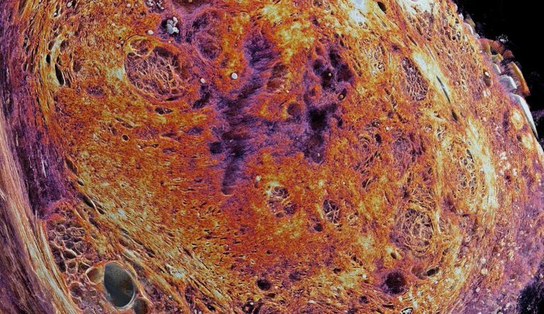
3D cinematic rendering of Hierarchical phase-contrast tomography (HiP-CT) depicting the resection of a human breast carcinoma (left). 3D cinematic rendering of Hierarchical phase-contrast tomography (HiP-CT) showing human pancreatic tissue with a neuroendocrine tumor (right) andhighlighting the vascularity of a human spleen.
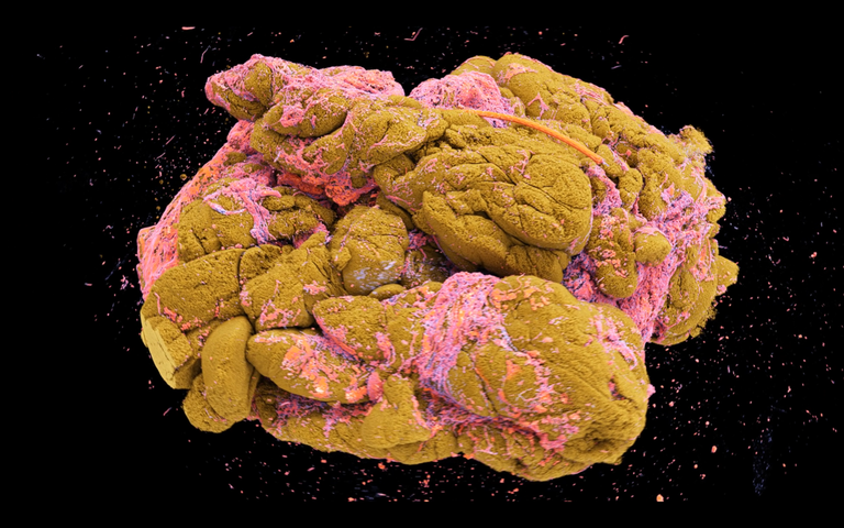
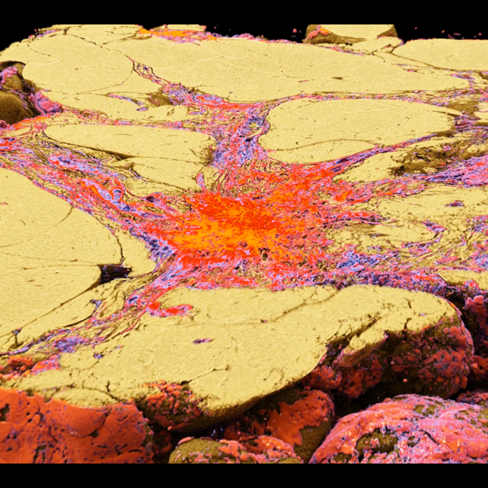
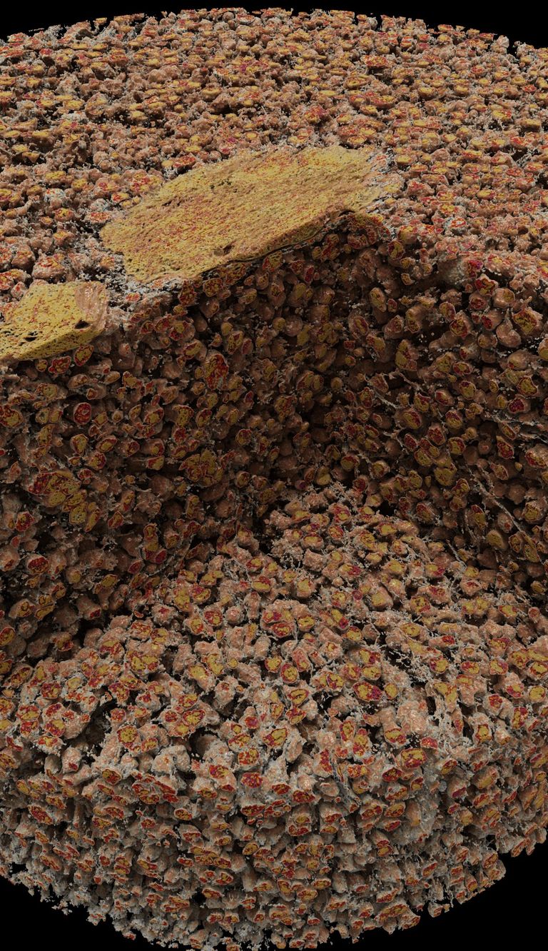
Breast cancer
Cinematic rendering of a synchrotron-based HiP-CT scan of invasive breast cancer provides a clear three-dimensional image of a five-centimetre piece of tissue removed from the breast of a breast cancer patient (see above). The fatty tissue and lobules (yellow) and the fibrous structure of the connective tissue (pink), along which the cancer spreads in the breast, are clearly visible. Siemens Healthineers AG kindly provided the cinematic rendering software.
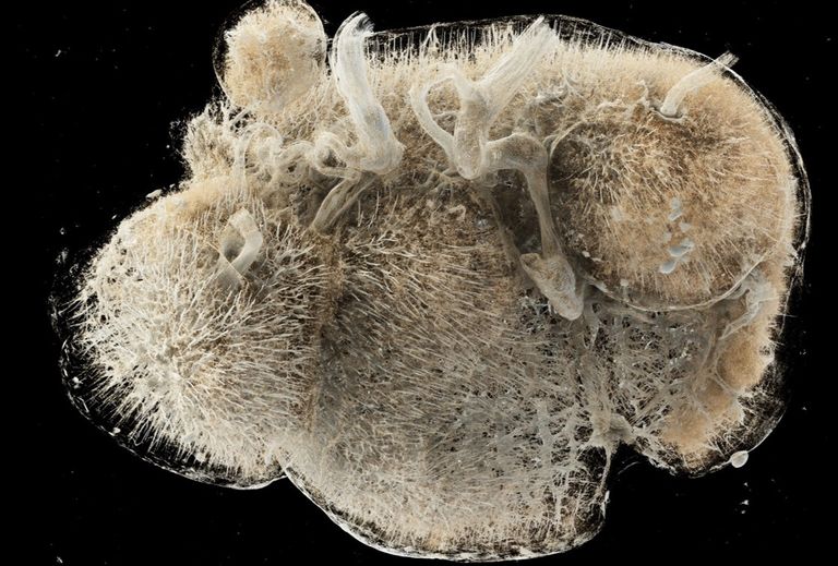
Bone marrow biopsy
Cinematic rendering of a bone marrow biopsy with MPN/MDS overlap depicting the fibrotic remodeling.
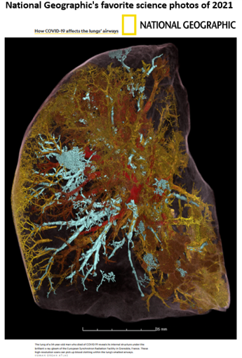
COVID-19 lung
HiP-CT scan of a COVID 19 lung was awarded as one of National Geographic's favorite science photos of 2021
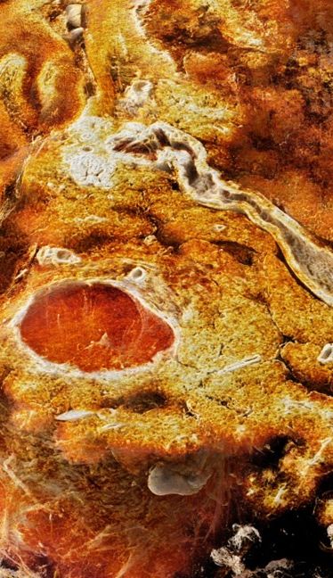
Prostate
HiP CT scans reveal the adenomyomatous hyperplasia in BPH.

Himalaya-Project
In cooperation with physicists, data science experts, and experienced mathematicians (Siemens Healthineers, German Cancer Research Center DKFZ, the Karlsruhe Institute of Technology (KIT)), machine learning and deep learning algorithms will be applied using clinically annotated, rendered synchrotron-radiation-based µCT datasets, whole-slide histological scans, and MRI scans (preoperative PI-RADS, and ex vivo scans) to set up convolutional neural networks with deep learning of biomedical features (3D tumor growth, tumor grading, a new 3D Gleason score, capsule infiltration, molecular pattern etc.).The superior integration and interpretation of these clinical and biological fully characterized data allows the implementation of AI and Deep Learning Algorithms for the optimization of prostate cancer screening tools, esp. MRI-based PI-RADS scoring, by automatically characterizing and quantifying the anatomies and abnormalities of prostate tissue and improving the diagnostic process. Moreover, the intimate integration of molecular tumor profiles with the clinical characteristics and 3D radiology data creates a unique opportunity for AI applications to improve tumor and metastasis detection, precise classification, and overall prognostication by applying AI-based Predictive Modelling of tumor growth and progression behavior.
www.himalaya-project.net


