

In the field of medicine, pathology is frequently referred to as the "pilot" discipline, given its pivotal role in diagnostics and therapeutic interventions. The examination of tissue and cells is essential for the accurate identification and classification of diseases, which forms the foundation for effective treatment. Microscope techniques are utilised to observe and document the various morphological changes associated with the disease.
Since the mid-19th century, when Rudolf Virchow co-founded the field of pathology, tissue diagnoses have been made to the present day on fine tissue sections using a simple light microscope, as we are sometimes still familiar with from our school biology lessons. In recent decades, however, the diagnostic precision of cancers has not only increased thanks to new possibilities in molecular biology and genetics, but considerable progress has also been made in biophysical imaging. The latest imaging techniques provide a broad range of possibilities for high-resolution three-dimensional tissue analysis. By acting as a virtual microscope, these techniques can bridge the gap between the macroscopic and microscopic pillars of radiology and pathology.

LUNG
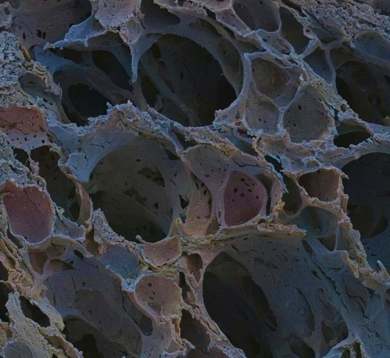
Healthy lung
Coloured scanning electron micrograph of a human lung with numerous lung baskets (alveoli).
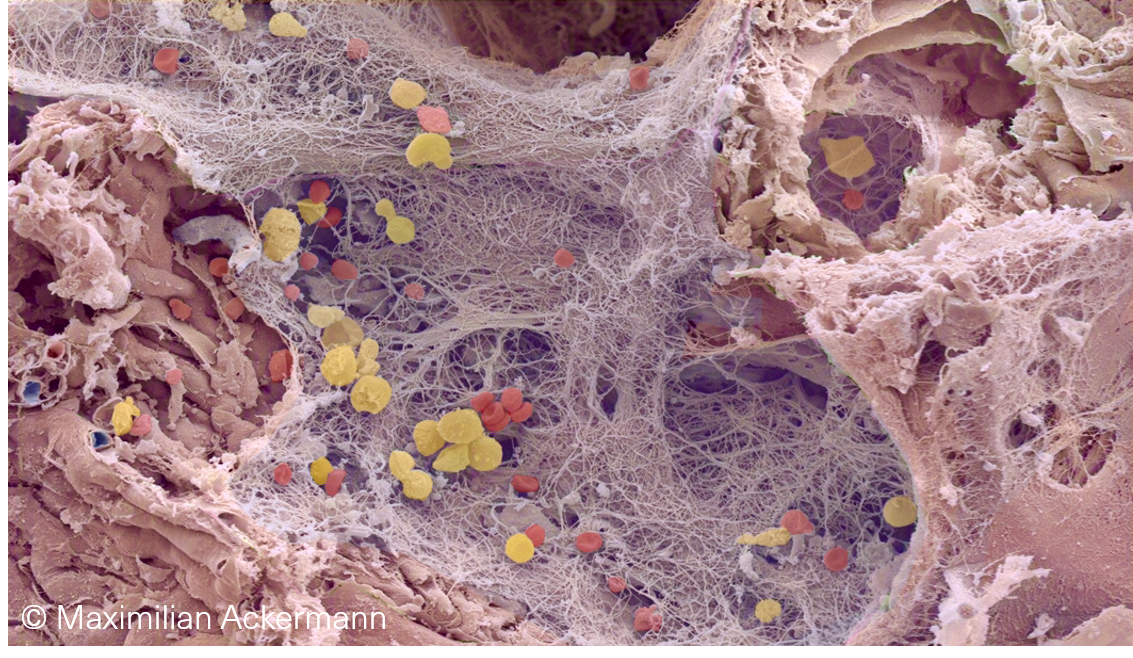
Lung fibrosis
Coloured scanning electron micrograph of a human lung with idiopathic lung fibrosis and scarring.
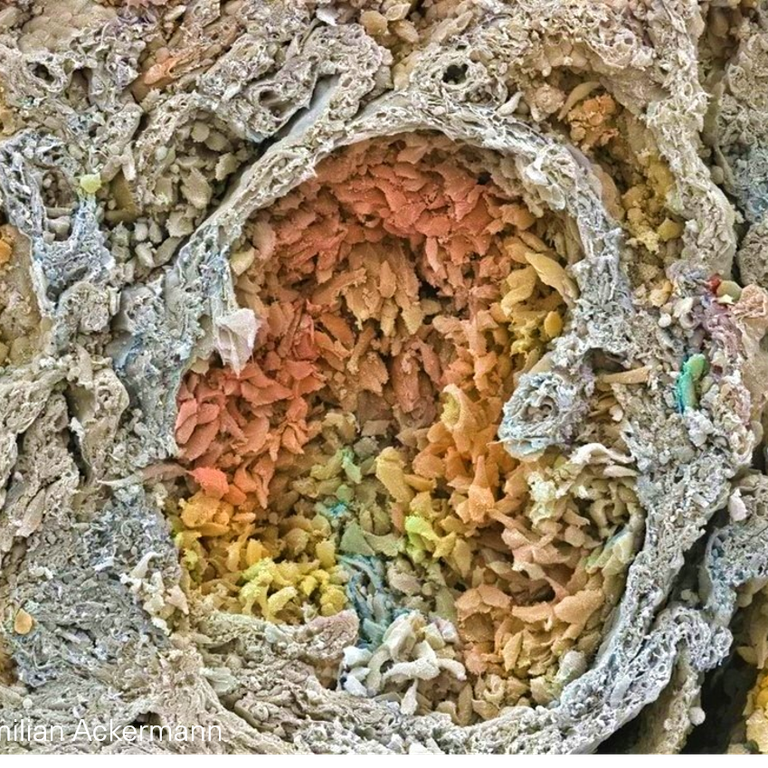
Lung carcinoma
Coloured scanning electron micrograph of a human pulmonary adenocarcinoma.
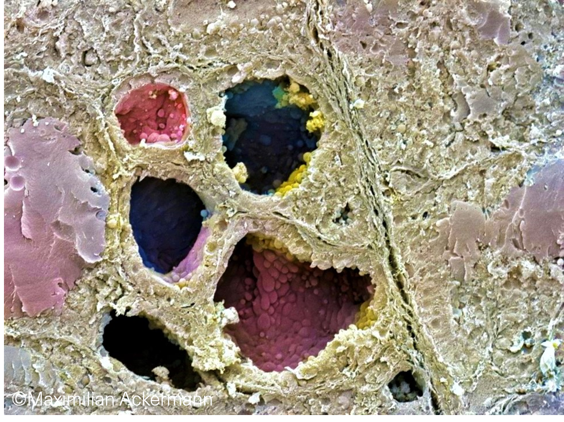
LYMPH NODE & BONE MARROW
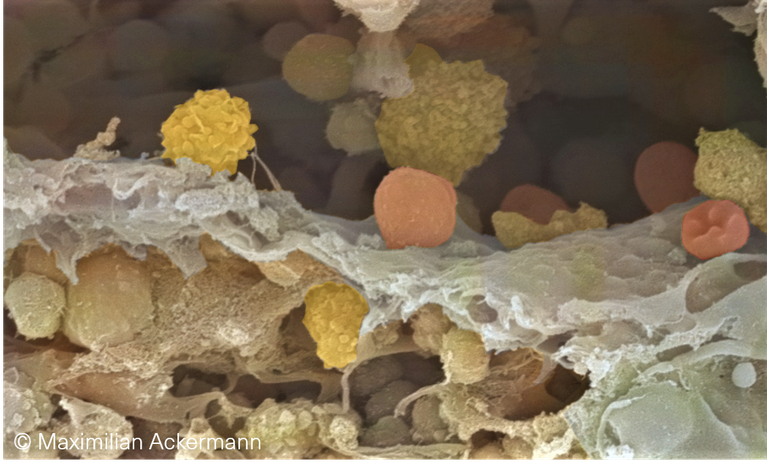
Coloured scanning electron micrograph of a human bone marrow sinus with the shedding of platelets.
Coloured scanning electron micrograph of a sinus in a human lymph node depicting
the transmigration and homing of lymphocytes (yellow) into the lymph node.
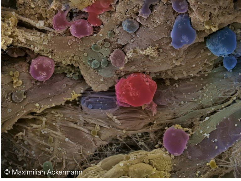
CARDIOVASCULAR DISEASES
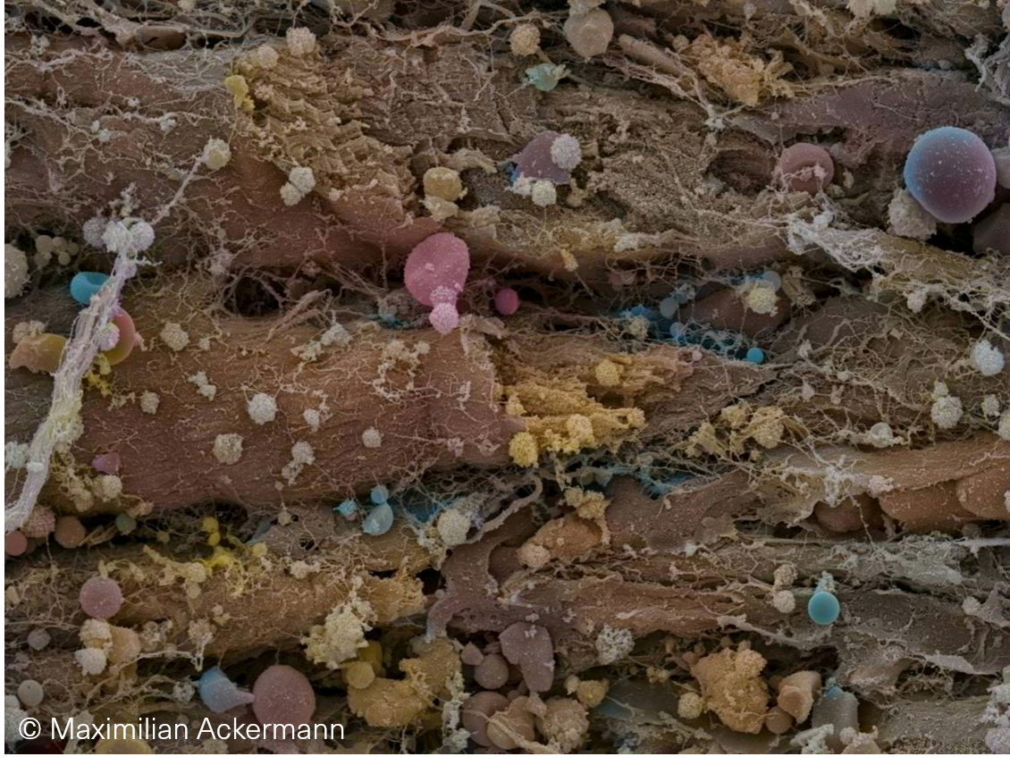
Coloured scanning electron micrograph of a human heart explant shows the dense infiltration of cardiomyocytes by lymphocytes in a heart with Parvovirus B19 myocarditis.
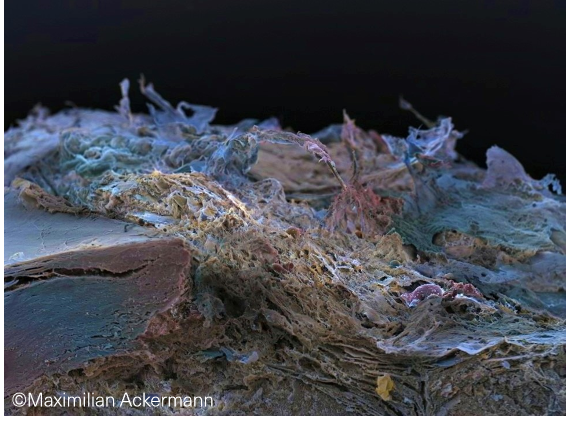
Coloured scanning electron micrograph of human aorta with artheroscelorosis, so called „coral reef artherosclerosis“, rock-hard calcifications with desctruction of the aortic wall.
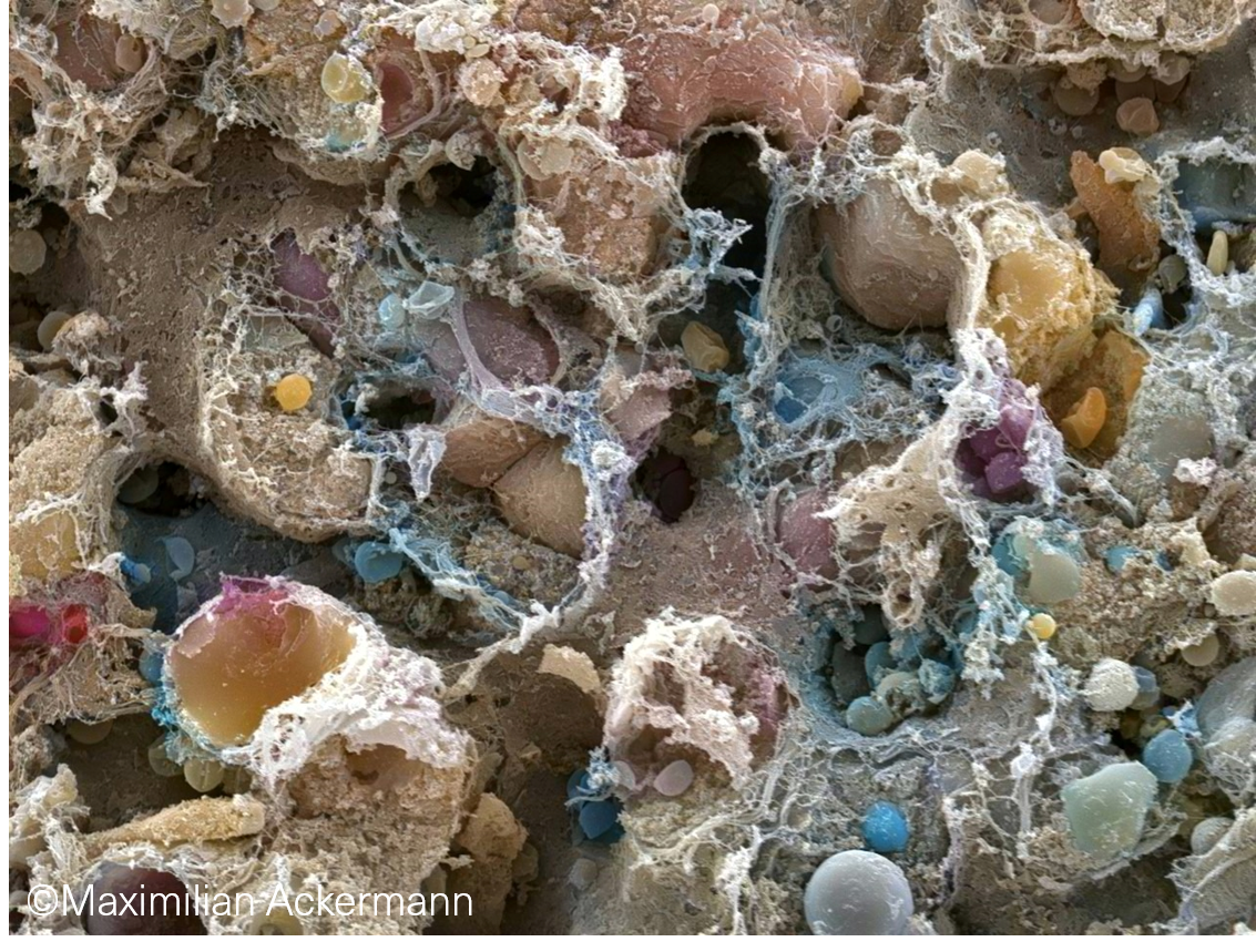
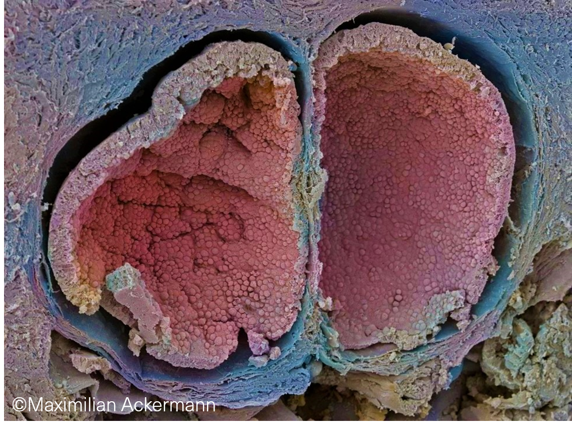
GASTROINTESTINAL TRACT
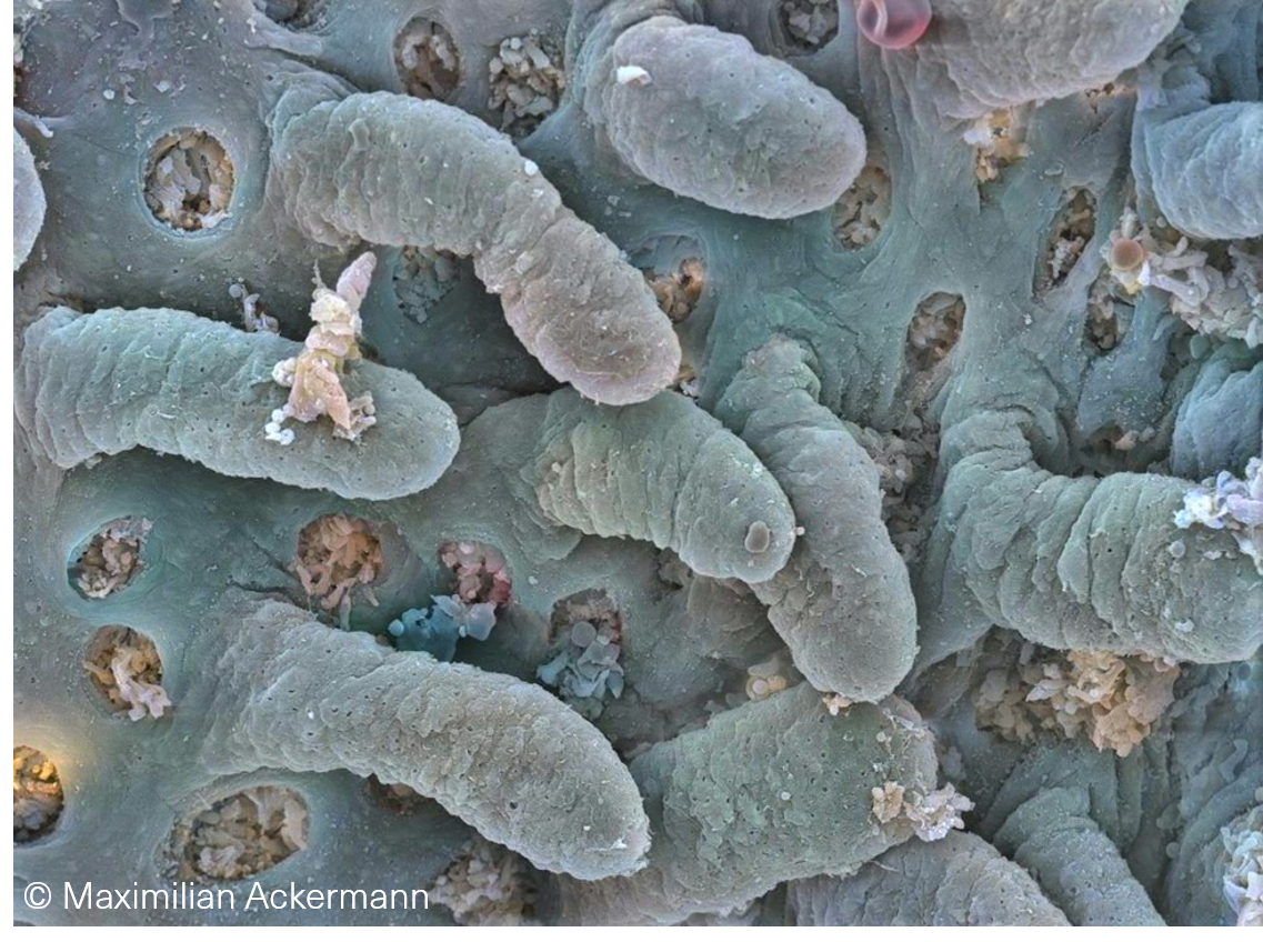
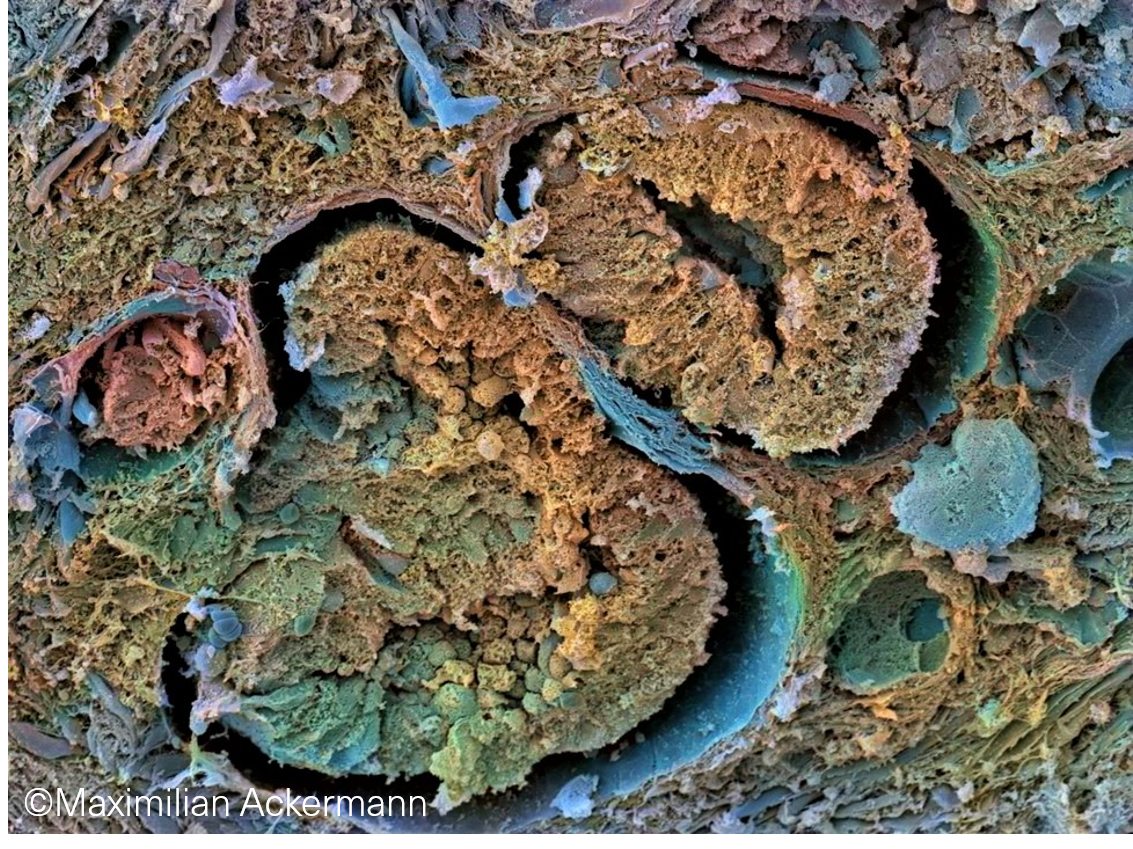
Coloured scanning electron micrograph of human duodenum depicts numerous villi protruding into the intestinal lumen.

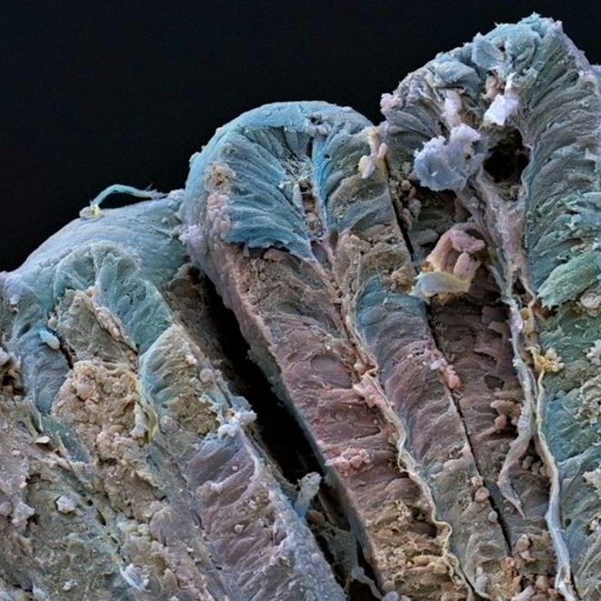
Colonic mucosa
Coloured scanning electron micrograph of
the surface of a human colonic crypt.
Colonic Polyp
Coloured scanning electron micrograph of
the surface of a human colonic polyp
showing pseudostratified and pencillate
nuclei extending to the surface.
Colon carcinoma
Coloured scanning electron micrograph of
the surface of a human colonic
adenocarcinoma showing the loss of
hierarchy and polarity and the infiltration
by lymphocytes (yellow).

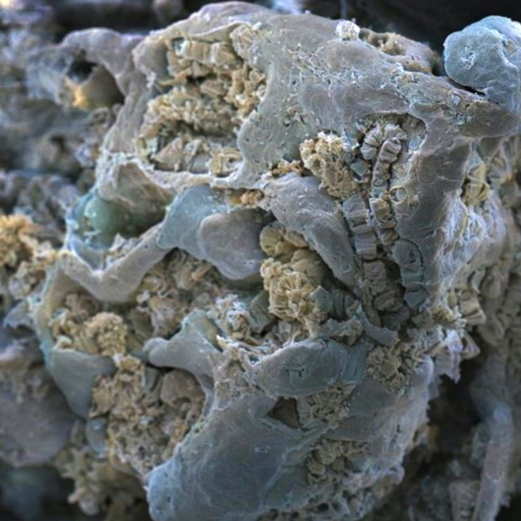
Coloured scanning electron micrograph of a human gallstone.
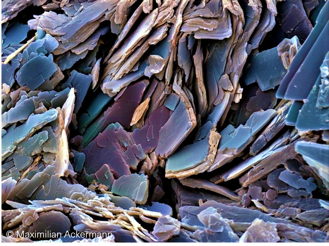
Coloured scanning electron micrograph of a human gallstone looks like gray slate.
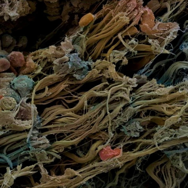
Coloured scanning electron micrograph of a human liver with the trabecular shape of liver cells (hepatocytes), liver sinusoids and the deposition of collagen bundles.
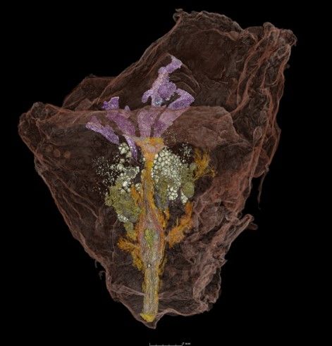
PROSTATE
The Himalaya Project (HIMALAYA: Holistic Imaging and Molecular Analysis in Life-Threatening Ailments," aims to improve prostate cancer diagnosis and risk assessment. To this end, we are using new, experimental imaging techniques that enable an unprecedented level of detail in prostate cancer imaging. One important technique is imaging the entire prostate, including the tumor and its surroundings, using synchrotron radiation from a circular particle accelerator as part of nanotomography. The high image quality, combined with histological examination and additional molecular testing, improves the interpretation of MRI images. This allows us to better understand prostate cancer and develop new diagnostic and therapeutic approaches.

Coloured scanning electron micrograph of a human prostate with prostate stones (calcium oxalate deposits).
Coloured scanning electron micrograph of a human benign prostatic hyperplasia (BPH) with expanding glands.
Coloured scanning electron micrograph of a human acinar adenocarcinoma of the prostate (Gleason pattern 3).

Coloured scanning electron micrograph of a human acinar adenocarcinoma of the prostate with a tertiary high grade pattern.
BRAIN
Coloured scanning electron micrograph of cerebral microvasculature shows the microvascular aneurysms and pompons indicating the vascular abnormalities in Alzheimer’s disease.

Coloured scanning electron micrograph of a human brain with COVID19 with microclots.

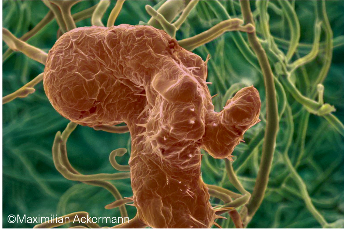
Coloured scanning electron micrograph of spheroid composed of glioblastoma cells.
TOOTH
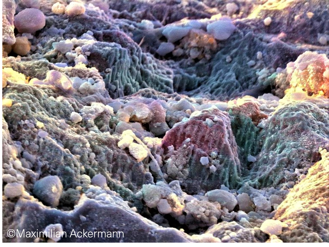
Coloured scanning electron micrograph of a human tooth surface with a carious lesion and exposed hydroxyapatite crystals.